Pleural Fluid - LDH:
Why Pleural Fluid LDH Test ?
CLINICAL INFORMATION
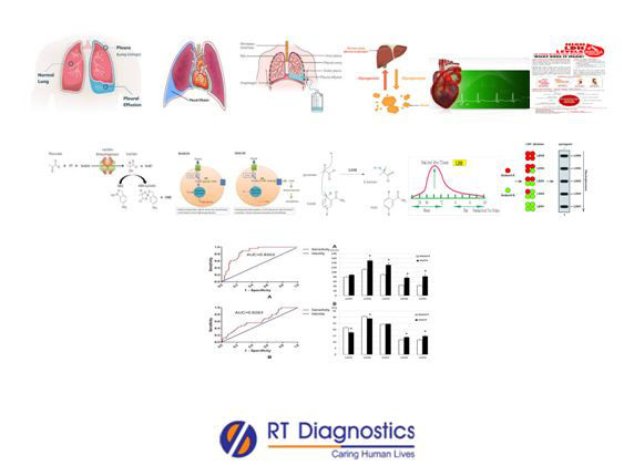
Pleura are a membrane lining the thoracic cavity (parietal pleura) and covering the lungs (visceral pleura). The parietal pleura folds back on itself at the root of the lung to become the visceral pleura. A small amount of space between the two pleurae and lubricating fluid at their contact points allow the lungs to expand. The pleura exude a thin fluid that keeps it moist and lubricated. Thus the pleura with two thin layers of tissues protect and cushion the lungs. The pleural fluid lubricates the pleural cavity so that the pleural tissue can slide against each other. Pleural fluid analysis is required in pathologies including chylous effusion, chyliform effusions, hemothorax, empyema, trapped lung, iatrogenic effusions, malignancy in lungs, certain cancers such as lymphoma, mesothelioma, and blood cancers (metastatic cancers), inflammation (pleuritis), infections, injuries, pulmonary embolism etc. Indication for pleural fluid for analysis arise when clinical manifestations with signs and symptom of chest pain, dyspnea, orthopnea, non-productive cough, trouble breathing, fatigue, X-ray findings - fluid blunts the posterior costophrenic angle, blunting of lateral costophrenic angle, effusion causing opacity portions of the hemithorax causing a mediastinal shift to contralateral side etc, and thus the imaging studies like X-rays are required for the confirmation of pleural effusion (compared with Light’s criteria protocol).Untreated chronic cases may lead to complications such as collapsed lung, pulmonary edema etc. Pleural fluid testing involves certain tests such as visual examination, odour analysis, microscopic evaluation, cytology, markers of pleural inflammation (CRP, IL-6 induced by lipopolysaccharide, TNF-tumor marker, procalcitonin, ADA, tumour markers – CEA, CA 125, CA 15-3, CYFRA 21-1 i.e it is a cytokeratin tumour marker in non-small cell lung cancer, ANA in rheumatoid arthritis and SLE, RF titers), culture and sensitivity tests, acidity (pH paper), cell count, total protein, LDH, glucose, based on patient’s history the test may also include lipid profile, creatinine, amylase, NT-proBNP, imaging studies, examination and radiology etc. A pleural fluid test is performed to analyse for differential diagnosis for transudative effusion (i.e extra fluid leak into the pleural space - fluid accumulation is caused due to mostly non-infectious origin such as severe malnourishment or rarely by pathologies like hepatic fibrosis, heart failure eg congestive heart failure, cirrhosis etc - i.e occurs due to the change in oncotic pressure exerted by serum albumin) and exudative effusion (fluid accumulation is caused due to some pathological origin i.e due to mainly infectious microbes and other causes like cancers, kidney diseases, auto-immune diseases, pneumonia etc) for treatment. This differentiation is based on fluid albumin level i.e the Serum-Ascites Albumin Gradient (SAAG) calculation, which is serum albumin level minus the fluid albumin level will distinguish if it is a case of exudate or transudate. The most common causes of lung and pleural injuries include injuries of the chest (physical trauma), viral, fungal, and bacterial causes of pleural effusion due to Streptococcus milleri group, Streptococcus pneumonia, Streptococcal aureus etc and also autoimmune diseases, rheumatoid arthritis, tuberculosis etc. Symptoms of pleural effusion include chest pain, dry and non-productive cough, dyspnea, orthopnea etc. Thoracentesis is a procedure where the needle is inserted through the chest wall into the pleural space to remove fluid or air from around the lungs. Pleural fluid testing is used to help diagnose the cause of fluid build-up in the chest (pleural cavity) which includes fluid appearance, cell count and fluid protein, albumin or LD level. The common complication of thoracentesis is pneumothorax, fluid build-up in the lung, excessive bleeding while specimen collection, re-accumulation of fluid in the lung, infection at the site of thoracentesis, respiratory distress or breathing difficulties etc. Culture and Sensitivity – Pleural Fluid Specimen test: This test specimen of Pleural fluid examines the aspirated fluid sample specimen from the pleural space in case of suspected infection and/or to diagnose the cause of fluid build-up in the space of the chest wall (for differential diagnosis to differentiate between exudates and/or transudate). Inflammation of the pleura causing exudates include infections, bleeding (bleeding disorders, trauma, pulmonary embolism, lung diseases eg. Sarcoidosis) and other causes like heart surgery, heart or lung transplant, pancreatitis and/or abscess in the abdomen etc. The non-infectious causes behind pleural effusion also include transudate (due to altered levels in the protein i.e low protein levels in the blood and/or injury or inflammation to the pleura - an imbalance between the pressure of the liquid within the blood vessels, drives fluid out of blood vessel), due to congestive heart failure or cirrhosis, tuberculosis, sarcoidosis, lung cancer, metastatic cancer, lymphoma, mesothelioma, RA, SLE etc. Gram stain also called as Gram stain method used for staining to classify two groups of bacterial species such as gram-positive and gram-negative bacteria. Bacteria with gram-positive cell walls have thick peptidoglycan which retains the primary stain crystal violet and Lugol’s iodine solution is added to strengthen the bonds of the stain. While the gram-negative bacteria since they have a thinner bacterial cell wall allow the crystal violet to wash out with the addition of ethanol, hence their cell membrane are stained pink or red by the counterstain (safranin or fuchsine). In certain cases pleural decortication is performed by the surgeons to operate inside the pleural space to remove unhealthy cells (potentially dangerous inflammation). Moreover, pleurodesis may be required in patients with frequent pleural effusion. The pleurodesis procedure involves the placement of certain substances such as doxycycline into the pleural space that inflames the pleura and on healing the two layers merge together. In General, the enzymes within the cell (cytosol) are impermeable to cell membranes (hence these cellular enzymes cannot enter blood circulation easily) and thus these enzyme activities are very low compared to those within the cell. ‘lactate dehydrogenase is also known as LDH. This enzyme catalyzes the conversion of lactate to pyruvate. LDH is present in almost every cell in the body- including blood (serum and/or plasma), muscles, brain (CSF sample specimen collected by lumbar puncture also known as a spinal tap), kidneys, liver, lung (eg. empyema), pancreas etc. “Iso-Enzymes” are the multiple forms of the same enzyme that differ with physical properties but they have the same function (similar catalytic activity) eg- they bind to the same substrates (or in other words - They are different variants of the same enzyme having identical functions). Iso-enzymes are of utmost clinical interest since they can be used as molecular markers of tissue damage. The iso-forms of LDH are LDH-1: present in the heart, brain and RBCs, LDH-2: present in the RES – Reticulo-Endothelial System, LDH-3: present in the lungs, LDH-4: present in the kidney, placenta and the pancreas, LDH-5: present in the liver, striated muscles and the brain. LDH test measures the amount of LDH enzyme present in the blood and/or other body fluids. Hence this test looks for signs of damage to the body tissues (eg. Myocardial infarction) and also to know the extent of injury in case of heart, liver, kidney diseases etc. This test is a helpful tool in conditions like hemolytic and/or megaloblastic anaemia, prognosis in cancer treatment etc. The sample specimen is usually an intra-vascular fluid eg. Blood (serum/plasma) and/or body or bio-fluids. High levels of LDH are found in certain pathological conditions such as anaemia, liver diseases, muscle injury, kidney diseases, heart attack (myocardial infarction), pancreatitis, infections like infectious mononucleosis, encephalitis, meningitis etc, and certain cancer types leukaemia, lymphoma etc. Additional tests on pleural fluid other than culture and sensitivity include measurement of pleural fluid, estimation of biochemical parameters such as glucose, lactate, amylase, triglyceride, tumour markers such as CEA etc. Supporting tests other than LDH include Troponin-1 (the diagnosis of infarction has been largely superseded by this test) test. Supporting tests include other serum levels of skeletal muscle enzymes or proteins that serve as markers in pathological conditions for eg. Creatine kinase (CK), aldolase, myoglobin, troponin, aspartate aminotransferase, carbonic anhydrase CAIII. But in physiological conditions too for example in strenuous exercises – muscle injury mediated by apoptosis may occur triggered by increased oxidative stress and hence other markers may serve as an important tool like thio-barbituric acid- reactive substances, malondialdehyde, sulfhydryl groups, reduced glutathione, oxidized glutathione, super-oxide dismutase, catalase etc. Other supporting tests include CBC, albumin, glucose, ESR, D-dimer, CRP, enzymatic assays or colourimetric, HPLC, spectrophotometry, Chest X-Rays, CT scans, Gram stains, fungal tests, stool tests for cysts in parasitic infestation, AFB test and adenosine deaminase to detect tuberculosis etc.
General Instructions:
Sample Requirement: Specimen –Pleural Fluid. Test Preparation: None.
NOTE - Sample for specimen collections may vary based on the patient’s condition/cases according to the patient’s presenting complaints/signs or symptoms:
SPECIMEN REQUIREMENT (Special or Rare Cases) - As instructed and guided by Physician / Clinician / Pathologist / as per Laboratory’s requirements, according to procedures and protocols.
This Multi-Specialty Clinical Referral Laboratory RT DIAGNOSTICS provides precise and accurate tests with an extensive range of testing services to the medical centres to help in the diagnosis and identification of pathology in the test specimens for infectious diseases and also to evaluate the function of organ systems of the patient. It prevents further complications and helps to stabilize and restore health to near normalcy at the earliest without delay.