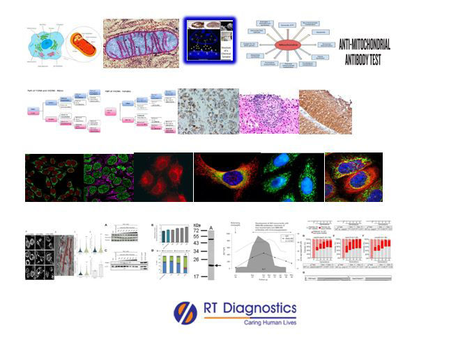
Mitochondrial Antibody:
Why Mitochondrial -Antibody Test?
CLINICAL INFORMATION
Mitochondria are membrane-bound cell organelles that generate most of the energy (ATPs – Adenosine Triphosphate) needed to power the cell’s biochemical reactions, hence they are called (energy currency or energy factory) “the powerhouse of the cell”. Hence mitochondria produce 90% of the energy our body needs to function. A mitochondrion is a cell organelle present in the cytoplasm of eukaryotic cells. Like chloroplasts, mitochondria are also considered prokaryotic in origin – since they have striking similarities to that of bacterial cells, as they have their own mitochondrial DNA to synthesize their own proteins required for their function. Hence a hypothesis suggests that the evolution of mitochondria could be endosymbiotic (descended from specialized purple non-sulfur bacteria). Most of the cell’s DNA are present in the nucleus of the cell, but the mitochondria have their own independent genome (the mitochondria genome encodes 37 genes) known as the mitochondrial DNAs (mDNA or mtDNA). These mitochondrial DNAs are highly susceptible to mutations (since it does not contain the DNA repair mechanism common to nuclear DNA. Moreover, mitochondria are the major site for the production of Reactive Oxygen Species (ROS) – also called free radicals, since they can release abundant free radicals (which might also be responsible for mutating the mitochondrial DNA itself but the presence of antioxidant proteins within the mitochondria scavenge and neutralise these free radicals thus preventing from its damage). Mitochondria divide by binary fission (like prokaryotes) and the mitochondria may replicate their DNA and divide in response to the energy needs of the cell (rather than in phase with the cell cycle). Mitochondria use oxygen and glucose to produce most of the cell’s energy known as Oxidative phosphorylation. The brain consumes a large amount of ATP used in most biochemical reactions of the neurons. Each neuron contains a huge amount of mitochondria within them. In neurons, the mitochondria are accumulated in the Presynaptic and postsynaptic locations (at the synaptic terminals where the regional ATPs in between the 2 neurons and cytosolic calcium levels modulate signal transductions) where a lot of energy is necessary. The function of mitochondria is to generate heat, synthesize ATPs or energy for the cells, cell singling, cell growth, cell differentiation, cell death – apoptosis, plays a role in calcium homeostasis, involves in the regulation of innate immunity, modulates stem cell regulation etc. Primary mitochondrial dysfunction can inhibit apoptosis (cell death), hence contributing to the development of cancers. Many conditions can lead to secondary mitochondrial dysfunction and affect other diseases including Alzheimer’s disease, muscular dystrophy, Lou Gehrig’s disease, diabetes, cancers etc. Mitochondrial disorders (often impacts multiple organ systems) can cause a vast array of health concerns including fatigue, weakness, metabolic strokes, seizures, cardiomyopathy, arrhythmias, developmental and cognitive disabilities, impaired vision, disorders of the liver, kidney, gastrointestinal tract, stunt growth, mitochondrial myopathy, Diabetes And Deafness (DAD), Leber’s Hereditary Optic Neuropathy (LHON), Leigh syndrome, subacute necrotizing encephalopathy, neuropathy, ataxia, retinitis pigmentosa, ptosis, myoneurogenic gastrointestinal encephalopathy, elevated levels of amino acids along with aminoaciduria (a sign of mitochondrial renal tubular dysfunction) and respiratory chain dysfunction etc. Autoimmune diseases associated with mitochondria are referred to as Anti-Mitochondrial Antibodies (AMAs). AMA is most often related to a condition known as Primary Biliary Cholangitis – PBC (previously known as primary biliary cirrhosis. PBC is caused by an auto-immune attack on the bile ducts in the liver and hence leads to scarring and liver failure in many cases. There is also a high risk for the incidence of liver cancer. Clinical manifestations of PBC include jaundice, fatigue, itchy skin, abdominal pain, edema, ascites, dry mouth and eyes, weight loss etc. Additional tests include creatine phosphokinase, creatine, carnitine, bilirubin, albumin, CRP, Anti-nuclear antibody tests, Anti-Smooth Muscle Antibodies (ASMA), genetic testing etc. Other tests include CBC, LFT (Liver Function Test), imaging studies, EKG etc.

General Instructions:
Sample Requirement: Specimen - Blood sample collected from the vein. Test Preparation: None.
NOTE - Sample for specimen collections may vary based on the patient’s condition/cases according to the patient’s presenting complaints/signs or symptoms:
SPECIMEN REQUIREMENT (Special or Rare Cases) - As instructed and guided by Physician / Clinician / Pathologist / as per Laboratory’s requirements, according to procedures and protocols.
This Multi-Specialty Clinical Referral Laboratory RT DIAGNOSTICS provides precise and accurate tests with an extensive range of testing services to the medical centres to help in the diagnosis and identification of pathology in the test specimens for infectious diseases and also to evaluate the function of organ systems of the patient. It prevents further complications and helps to stabilize and restore health to near normalcy at the earliest without delay.


